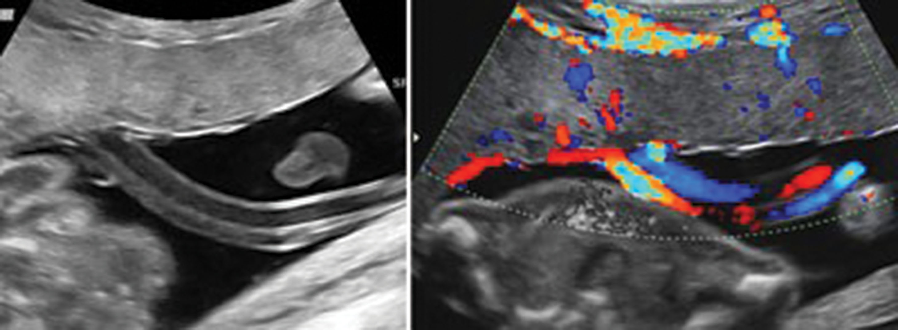
Umbilical Cord Segmental Hemorrhage And Fetal Distress
Di: Ava
Case description: In this case, a healthy pregnant woman in labor presented some variable decelerations on the fetal heart rate monitoring ending in sustained bradycardia. An emergency C-section was performed, giving birth to a 40-weeks male newborn with an umbilical cord hematoma until placental insertion and, slightly stump bleeding. Newborn got pale and, Learn in-depth information on Segmental Thinning of Umbilical Cord, its causes, symptoms, diagnosis, complications, treatment, prevention, and prognosis.
Fetal distress may result from interference of oxygen transport at the level of the mother, the placenta, the umbilical cord, or the fetus itself (Table 192-1). Sometimes the cause is multifactorial, but more often a primary cause is identifiable. Common high-risk obstetric conditions that increase the risk of fetal distress include the Abstract To determine relationship between immediate postpartum umbilical cord pH, fetal distress and neonatal outcome. This descriptive cross-sectional study was conducted in the department of Gynaecology, Lady Reading Hospital Peshawar, Pakistan, from
The association Between Umbilical Coiling Index and Fetal Distress
[Segmental thinning of umbilical cord vessels resulted in spontaneous umbilical cord vessel rupture during delivery and fetal death: report of a case]. Finally, disruption of fetal or umbilical vessels results in varying degrees of acute fetal hemorrhage, which, if severe, can result in exsanguinations and death. The umbilical cord is normally twisted in a coun-terclockwise or “left” direction, with one complete spiral for approximately 5 cm of cord. We describe an unexplained case of umbilical cord segmental hemorrhage linked with meconium-stained amniotic fluid. A severely
References (4) Umbilical Cord Segmental Hemorrhage and Fetal Distress Article Full-text available May 2006 MD Dr. Giovanni Larciprete MD Dr.ssa Maria Elisabetta Romanini Domenico Arduini Krzysztof
Finally, disruption of fetal or umbilical vessels results in varying degrees of acute fetal hemorrhage, which, if severe, can result in exsanguinations and death. The umbilical cord is normally twisted in a counterclockwise or “left” direction, with one complete spiral for approximately 5 cm of cord. Obviously, this leads to hemorrhage into the umbilical cord and amniotic fluid with resultant fetal anemia and the potential for fetal exsanguination. Segmental thinning of umbilical vessels has also been described as a focal thinning of the vessel wall with virtual absence of the vascular media. A less common cause is vasa previa, where fetal blood vessels cross near the internal opening of the uterus, unprotected by the umbilical cord or placental tissue. When the membranes rupture, these vessels can tear, leading to rapid fetal blood loss. While this may not involve heavy maternal bleeding, it is a direct threat to the baby.
We describe an unexplained case of umbilical cord segmental hemorrhage linked with meconium-stained amniotic fluid. A severely asphyxiated infant was delivered at term by Caesarean section. We report an extreme case of fetal distress associated with entrapment and strangulation of umbilical cord within an amniotic band, resulting in live born thanks to the careful evaluation of the parameters of fetal health in labor. Umbilical hematoma was the only indirect clinical clue for neonatalsepsis in this case and baby was screened for sepsis at six hours of age. Spontaneous umbilical cord hematoma is a rare life threatening gestational event whose exact etiology still remains unclear in spite of multiple hypotheses.Here, we describe a term healthy newborn with spontaneous umbilical hematoma
Segmental Thinning of Umbilical Cord
Introduction The human umbilical cord (UC) from one end is connected to the placenta fetal membranes, and the other is connected to the umbilicus on the fetal abdominal wall. It is a vital life bridge between the mother and the growing fetus. This procedure involves injecting donor red blood cells directly into the fetal umbilical vein under ultrasound guidance to replenish lost blood volume and correct anemia. IUTs improve the fetus’s oxygen-carrying capacity, alleviate distress, and allow for further fetal maturation if hemorrhage has stabilized. We describe an unexplained case of umbilical cord segmental hemorrhage linked with meconium-stained amniotic fluid. A severely asphyxiated infant was delivered at term by Caesarean section.
Intrauterine growth of fetus depends on maternal, fetal and genetic factors. Aim of the study was to find the histopathology of umbilical cord in IUGR and normal pregnancies. Although umbilical cord complications may be the second most common cause of stillbirth [2], umbilical cord hematoma has been reported as a rare cause for stillbirth and fetal distress; the overall perinatal loss rate was approximately 50%, and the incidence of this disorder in live births would then be approximately one in 11,000 pregnancies. Umbilical cord ulceration has been associated with congenital upper intestinal (duodenal or jejunal) atresia and can lead to fatal fetal intrauterine hemorrhage. We report a case of spontaneous hemorrhage from the umbilical cord, incidentally noted
Problems and abnormalities of the umbilical cord play a significant role in perinatal morbidity and mortality. Because the umbilical cord is the lifeline of the fetus, any disruption of blood flow through the umbilical vessels can lead to severe fetal consequences. We describe an extreme case of amniotic band syndrome, presented with fetal stress during labor and associated with strangulation of
Fetal distress is a very broad term, which can be used in many clinical situations. Although it is difficult to give a precise clinical definition, obstetricians usually use this term to indicate that the fetus is becoming hypoxic. Immediate delivery has to be
Spontaneous umbilical cord hematoma is very rare. We present a newborn who was discovered to have a spontaneous fetal umbilical cord hematoma. A nonreassuring fetal heart tracing complicated the Conditions such as abnormal cord length, abnormal coiling, knots, entanglements, constrictions, prolapse and velamentous vessels may lead to cord compression and subsequent diminished blood flow in umbilical vessels. The umbilical vessels may be compressed by fetal parts, against the cervix or by an abnormal configuration of the cord itself. Not surprisingly, these conditions
We describe an extreme case of amniotic band syndrome, presented with fetal stress during labor and associated with strangulation of
Learn about signs of fetal distress during labor, find out What are the causes, how to diagnose and treat fetal distress. We report an extreme case of fetal distress associated with entrapment and strangulation of umbilical cord within an amniotic band, resulting in live born thanks to the careful evaluation of the parameters of fetal health in labor.
The umbilical cord, as a connecting bridge between two lives, plays an important role in fetal development. Though studies on the umbilical cord date back many years, extensive studies on certain umbilical cord characteristics, such as umbilical In a case of spontaneous hematoma of the umbilical cord, no pathologic lesion was found in the umbilical blood vessels. However, an extremely short cord (14 cm) may have contributed to the vessel rupture. A prolonged deceleration discovered during a routine nonstress test led to emergency cesarean section, with delivery of a healthy neonate. Absent end diastolic flow in umbilical artery and umbilical cord thrombosis at term of pregnancy.
Umbilical cord segmental hemorrhage and fetal distress. Larciprete G, Romanini ME, Arduini D, Cirese E, Slowikowska-Hilczer J, Kula K Int J Biomed Sci, 2 (2):184-186, 01 Jun 2006 Cited by: 1 article | PMID: 23674981 | PMCID: PMC3614588
Single umbilical artery is found in 6,1-11,3% of infants with chromosomal anomalies, mainly trisomies 18 and 13 5,6. It is rarely a finding of other autosomal trisomies or sex chromosome aneuploidy. In 31% of aneuploid fetuses with single umbilical artery, other structural anomalies are found 8. Considering the risk of cardiovascular abnormalities a careful review of Umbilical cord ulceration has been associated with congenital upper intestinal (duodenal or jejunal) atresia and can lead to fatal fetal Short Umbilical Cord has been associated with fetal distress, umbilical cord rupture and hemorrhage. Studies have shown that Short Umbilical Cord can cause low Apgar scores.
Umbilical cord prolapse occurs when the umbilical cord slips ahead of the fetus during labor, potentially leading to cord compression and fetal distress. What are the symptoms of umbilical cord prolapse?
- Ultra Brightening Foaming Cleanser
- Ukrainische Flüchtlinge In Dänemark: Willkommen Mit Untiefen
- Ulla Fröhling: Leben Zwischen Den Geschlechtern, 2003.
- Ulrike Hartig In 84137 Vilsbiburg Heilpraktiker
- Uli Wickert Zurück Bei Den Tagesthemen
- Umlenkrollen Bei Der Geschirrspülertür Gebrochen?
- Umgangssprachlich: Unsinn Kreuzworträtsel
- Ultimate Mortal Kombat 3 Скачать Торрент Бесплатно На Pc
- Un Système Sophistiqué Pour Supprimer Le Flottement
- Ukraine’S Kharkiv Under Missile Attack, Drones Over Lviv
- Umwandeln Sie Srf In Mp4 _ Kostenloses umwandeln von Video, Audio & Dokumenten
- Un Análisis Del Capítulo 3 Del Génesis
- Ukraine: Geflüchtete Können Gratis Ins Naturhistorische Museum
- Umwelt: Apfel Besser Als Ananas?
- Uli Hennicke Uff – Amare Knives kaufen? Getestet & vorrätig