Mri Atlas Of Ms Lesions : Mri atlas of ms lesions 1st ed.2008
Di: Ava
数据介绍数据集信息 ATLAS v2.0 (Anatomical Tracings of Lesions After Stroke) 是一个从 MR T1 加权 (T1W) 单模态图像中对脑中风病灶区域进行分割的数据集,并作为 MICCAI ISLES 2022 挑战赛的一部分。T1W MRI The PI-RADS assessment categories are based on the findings of multiparametric MRI, which is a combination of T2-weighted (T2W), diffusion weighted imaging (DWI) and dynamic contrast-enhanced (DCE) imaging. It is an accurate tool in the detection of clinically significant prostate cancer. In PI-RADS v2.1 clinically significant cancer is defined on pathology
Mri atlas of ms lesions 1st ed.2008
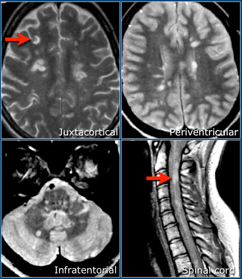
Multiple sclerosis is the most common chronic inflammatory disease of myelin with interspersed lesions in the white matter of the central nervous system. Magnetic resonance imaging (MRI) plays a key role in the diagnosis and monitoring of white matter diseases. This article focuses on key findings in multiple sclerosis as detected by MRI. MRI Atlas of MS Lesions M. A. Sahraian, E.-W. Radue MRI Atlas of MS Lesions With the collaboration of A. Gass, S. Haller, L. Kappos, J. Kesselring, J.-I. Kira, K. Weier 123 Prof. Dr. med. Mohammad Ali Sahraian Assistant Professor of Neurology Department of Neurology Tehran University of Medical Sciences Hassan Abad Square Sina Hospital Tehran-Iran

The images have been selected out of thousands of MRI images, and we hope that this MRI Atlas of MS Lesions, which is accompanied by a learning CD, provides valuable tools for clinicians and radiologists who are interested in MS for a better depiction of lesions, avoiding pitfalls, and demonstrating dissemination in space and time by MRI. MRI has become the main paraclinical test in the diagnosis and management of multiple sclerosis. We have demonstrated more than 400 pictures of different typical and atypical MS lesions in this atlas. Each image has a teaching point. New diagnostic criteria and differential diagnosis have been discussed.
Abstract MRI has become the main paraclinical test in the diagnosis and management of multiple sclerosis. We have demonstrated more than 400 pictures of different typical and atypical MS lesions in this atlas. Each image has a teaching point. New diagnostic criteria and differential diagnosis are discussed and the book is supported by a teaching DVD where the reader can Publicationdate 2021-12-01 This article is an updated version of the 2013 article and focusses on the role of MRI in the diagnosis of Multiple Sclerosis. We will discuss the following subjects: Typical findings in MS Role of MR in the McDonald criteria of MS How to differentiate MS lesions from other white matter diseases The importance of the a priori chance for the differential
Anatomical Tracings of Lesions After Stroke (ATLAS) R2.0 Overview Accurate lesion segmentation is critical for stroke rehabilitation research for both quantification of lesion burden and accurate image processing. Current automated lesion segmentation methods for T1-weighted MRIs lack the accuracy to be used reliably in research. MRI has become the main paraclinical test in the diagnosis and management of multiple sclerosis. More than 400 pictures of different typical and atypical MS lesions are demonstrated in this atlas. Each image has a teaching point. New diagnostic criteria and differential diagnosis are discussed. Daha fazla göster ISBN-10 3540713719 ISBN-13 978 Multiple hyperintense lesions on T2- and PD-weighted sequences are the characteristic magnetic resonance imaging (MRI) appearance of multiple sclerosis (MS). The majority of the lesions are small, although they can occasionally measure several centimeters in diameter. Focal MS lesions are usually round or oval in shape and relatively well circumscribed.
An MRI is the gold standard for detecting MS lesions, although some lesions do not show up on MRI scans — for example, some may be too small to be clearly seen through this technique. Monografie Neue Methoden der Computerunterstützung in der MRT-Diagnostik bei Multipler Sklerose Monografie Spatio-temporal segmentation and characterization of active multiple sclerosis lesions in serial MRI data Monografie Häufigkeit und Ursachen von Marklagerveränderungen im MRT bei pädiatrischen Patienten Monografie Automatic segmentation of multiple sclerosis (MS) lesions in brain MRI has been widely investigated in recent years with the goal of helping MS diagnosis and patient follow-up. However, the performance of most of the algorithms still falls far below expert expectations. In this paper, we review the main approaches to automated MS lesion segmentation. The main
New data emphasize early diagnosis and treatment with available disease-modifying therapies in MS (Rieckmam 2005), but establishing the diagnosis of MS is not always a straightforward process, and many other inflammatory and
- Anatomical Tracings of Lesions After Stroke
- ATLAS v2.0 数据集介绍
- Radue E.W., Sahraian M.A. MRI Atlas of MS Lesions
- Gadolinium Enhancing Lesions in Multiple Sclerosis
A. Lesions adjacent to lateral ventricle (Dawson’s fingers). MRI from a patient with early MS shows a few Dawson’s fingers on sagittal fluid-attenuated inversion recovery (FLAIR) image. Multiple sclerosis (MS) is a progressive neurological disorder marked by demyelinating lesions in the central nervous system. While MRI is essential for MS diagnosis, manual lesion segmentation is time-consuming and prone to variability. This study develops an automated deep learning (DL) approach for MS lesion segmentation using 3D-FLAIR MRI. MRI has become the main paraclinical test in the diagnosis and management of multiple sclerosis. We have demonstrated more than 400 pictures of different typical and atypical MS lesions in this atlas. Each image has a teaching point. New diagnostic criteria and differential diagnosis have been discussed.
The key question, then, is whether a patient’s MRI shows MS-like lesions. To answer this all-important question, I propose a systematic, checklist-based approach to reviewing brain MRI and have developed The MS Lesion Checklist based on my clinical experience and extensive literature review. It is not yet validated.
Mentioning: 5 – PrefaceMagnetic resonance imaging (MRI) has greatly increased our understanding about multiple sclerosis (MS) during the last two decades, and is now considered to be the imaging of choice for diagnosis and in vivo monitoring of the disease.New diagnostic criteria allow us to demonstrate dissemination of MS pathology in space and time by MRI, thus
Read & Download PDF MRI Atlas of MS Lesions by Mohammad Ali Sahraian, Ernst-Wilhelm Radue (auth.), Ernst-Wilhelm Radü, Mohammad Ali Sahraian (eds.), Update the latest version with high-quality. Try NOW! Dissemination in time: Simultaneous presence of enhancing and non-enhancing lesion on MRI, new T2/FLAIR hyperintense lesion on follow up MRI, or development of new clinical symptoms.
3/5 Sagittal FLAIR MRI from a patient with AQP4+ NMOSD demonstrating a marbled lesion in the corpus callosum following the ependymal lining (arrow). In recent years, there have been multiple works of literature reviewing methods for automatically segmenting multiple sclerosis (MS) lesions. However, there is no literature systematically and individually review deep learning-based MS lesion segmentation methods. Although the previous review also
In this blog post, we are going to share a free PDF download of MRI Atlas of MS Lesions PDF using direct links. In order to ensure that user-safety is not T1 black holes are hypointense lesions that may be seen on T1 weighted imaging in patients with multiple sclerosis, and indicates the chronic stage with white matter destruction, axonal loss and irreversible clinical outcome. The present book aims at demonstrating MS lesions in diferent sequences of conventional MRI, and shows examples of typical and atypi-cal lesions. The main idea for collecting the im-ages in this atlas is to show the diversity of MS lesions in diferent sequences of conventional MRI. There is a summarized introduction at the beginning of each chapter, followed by selected images in
Introduction Multiple sclerosis (MS) is an inflammatory demyelinating disease that the parts of the nervous system through the lesions generated in the white matter of the brain. It brings about disabilities in different organs of the body such as eyes and muscles. Early detection of MS and estimation of its progression are critical for optimal treatment of the disease.
MRI atlas of MS lesions by M Sahraian 0 Ratings 0Want to read 0Currently reading 0Have read
MRI Atlas of MS Lesions.pdf للمؤلف: M. A. Sahraian , E.-W. Radue تحميل كتاب MRI Atlas of MS Lesions.pdf رابط مباشر Magnetic resonance imaging (MRI) has greatly increased our understanding about multiple sclerosis (MS) during the last two decades, and is now considered to be the imaging of choice for diagnosis and in vivo monitoring of the disease
MRI has become the main paraclinical test in the diagnosis and management of multiple sclerosis. We have demonstrated more than 400 pictures of different typical and atypical MS lesions in this atlas. Each image has a teaching point. New diagnostic criteria and differential diagnosis have been discussed. MRI has become the main paraclinical test in the diagnosis and management of multiple sclerosis. We have demonstrated more than 400 pictures of different typical and atypical MS lesions in this atlas. B. Axial FLAIR MRI from a patient with AQP4+ NMOSD demonstrating an “arch bridge” lesion in the splenium of the corpus callosum (arrow; Image courtesy of Dr. Carlos Sollero). C. Coronal T1 postgadolinium MRI in a patient with AQP4+ NMOSD demonstrating an enhancing linear brainstem lesion in the right cerebral peduncle.
MRI has become the main paraclinical test in the diagnosis and management of multiple sclerosis. We have demonstrated more than 400 pictures of different typical and atypical MS lesions in this atlas. Each image has a teaching point. New diagnostic criteria and differential diagnosis have been discussed. 1/4 A. Brain MRI from Mr. L’s initial hospitalization with increased signal intensity in the right temporal lobe B. Brain MRI with enhancement of the temporal lesion (small arrow) with leptomeningeal enhancement (large arrow).
- Mountainbike Touren In Bad Berneck Im Fichtelgebirge
- Muay Thai Fulda Boxen??? : Kampfsportschule Düsseldorf Ratingen
- Mr. C Dj Casper Creator Of The Cha Cha Slide
- Msc Lauren Current Position – MSC LAUREN Position / Standort
- Muc-Off Wet Lube Ab € 17,99 _ Muc-Off E-Bike Wet Lube 50ml
- Moving Up Lyrics By Dannii Minogue
- Mountain’S Goat Diy-Set For Signature Drink Aufguss
- Ms Schwaben Im Art-Déco-Stil Umgebaut
- Msmq And Logging | Asynchronous Python Logging Using MSMQ
- Muenzenmaier, Antje Internisten Selm
- Mr. Potato’S Awesome Fitness Tips
- Movie: 365 Days Movie Official Trailer
- Ms Service In Großenseebach : Yener Sentürk Taxi Null Zwei Zwei, Großenseebach
- Msi Katana Gf66 12 Ab 849,00 €
- Mucaj Coiffeur Im Westend Elvana In Frankfurt Am Main