Ligaments Of The Knee Joint : Anatomy of the Knee Joint
Di: Ava
Knee anatomy is incredibly complex, and problems with any part of the knee anatomy, including the bones, cartilage, muscles, ligaments and tendons, can cause pain. The knee joint is stabilized by several ligaments, which are strong bands of connective tissue that connect bones to other bones. These ligaments play a What are knee ligaments? There are 4 major ligaments in the knee. Ligaments are elastic bands of tissue that connect bones to each other and provide stability and strength to the joint. The
This type of fracture is traditionally associated with tears of the anterior cruciate ligament of the knee (ACL), but increasingly also with the anterolateral ligament described
Anatomy of the Knee Joint
Printed publication (book, brochure, journal, etc.) Slide Presentation (Non-web or authenticated login if Web) Publishing in an Elsevier journal or book (print and/or electronic) Student Lo-res variable meniscofemoral ligaments originate from the posterior horn of the lateral meniscus and insert into the substance of the PCL. These include
The transverse or (anterior) meniscomeniscal ligament is a ligament in the knee joint that connects the anterior convex margin of the lateral meniscus to the anterior end of the medial
Read more about the four main ligaments of the knee, such as the anterior cruciate ligament (ACL) and the posterior cruciate ligament (PCL). The joint capsule works synergistically with the ligaments and tendons around the knee, such as the anterior and posterior cruciate ligaments, to maintain joint integrity during
In this video Prof Bellemans explains which 5 ligaments stabilize the knee, how they work, and how they may get damaged, stretched or tear.00:00 anatomy of t The knee is the largest joint in the human body. It is a compound joint. Not only is it where the femur, or thigh bone, meets the tibia, or shin bone – at th
On the medial aspect of the knee joint, we can also visualize two small ligaments that represent the deep layers of the tibial collateral ligament which attach to the medial Explore knee anatomy with a complete guide to parts, names, functions & diagram. Learn joint structure, ligaments & muscles for better The knee anatomy is a complex hinge joint that flexes, extends, and twists slightly from side to side. It is responsible for weight bearing and movement. The knee consists of bones,
Functional Anatomy of the Knee
- The 5 knee stabilizing ligaments
- Applied anatomy of the knee
- Functional Anatomy of the Knee
Function The coronary ligaments function to connect parts of the outside, inferior edges of the medial and lateral menisci to the joint capsule of the knee. The medial meniscus also has firm
On the injuries to the ligaments of the knee joint: a clinical study. 1938Clin Orthop Relat Res. 2007 Jan:454:17-22; discussion 14. doi: 10.1097/BLO.0b013e31802c7915. Background Our study aims to examine stress–strain data of the four major knee ligaments—the anterior cruciate ligament (ACL), the posterior cruciate ligament (PCL), the
Together with the posterior cruciate ligament (PCL), the ACL guides the instantaneous center of rotation of the knee, therefore controlling joint kinematics. While the anteromedial bundle is the The ligaments provide stability during loading while the muscles around the knee have a secondary role in stabilising this joint. If these structures are compromised, there may be A model of the right knee showing some of the key bones, ligaments and tendons. – Knee Anatomy Model – 3D model by University of Dundee, CAHID
- Anatomy and Biomechanics of the Collateral Ligaments of the Knee
- Ligaments of the Knee Joint
- Knee Joint Anatomy: Overview, Gross Anatomy, Natural Variants
- Normal Anatomy and Biomechanics of the Knee
- Medial Patellofemoral Ligament
Knee Anatomy Review The knee joint involves a complex arrangement of muscles, ligaments, nerves, and bones that work together to form the knee joint proper. The primary movements of The knee joint is also strengthened by the intracapsular structures, which include the cruciate ligaments and menisci. The two cruciate ligaments are located within the fibrous joint capsule The articular capsule of the knee joint is the wide and lax joint capsule of the knee. It is thin in front and at the side, and contains the patella, ligaments, menisci, and bursae of the knee. [1]
Comprehensive knowledge of medial knee joint biomechanics is valuable for the assessment of which injured structures should be repaired or reconstructed. An understanding The cruciate ligaments are the nucleus of knee joint kinematics (We. Müller 1977). In addition, they are the primary restraints to anterior-posterior translation of the tibia (Butler et al. 1980).
The Medial Patellofemoral Ligament (MPFL) is an hour-glass shaped ligament made of bands of retinacular tissue. The MPFL plays a significant role in the stabilization of the medial aspect of CHAPTER 20 The Knee CHAPTER OBJECTIVES At the completion of this chapter, the reader will be able to: Describe the anatomy of the joint, ligaments, muscles, and It’s actually attached medially to the tibial collateral ligament or sort of blended with the fibers of the tibial collateral ligament and also to the capsule of the joint of the knee.
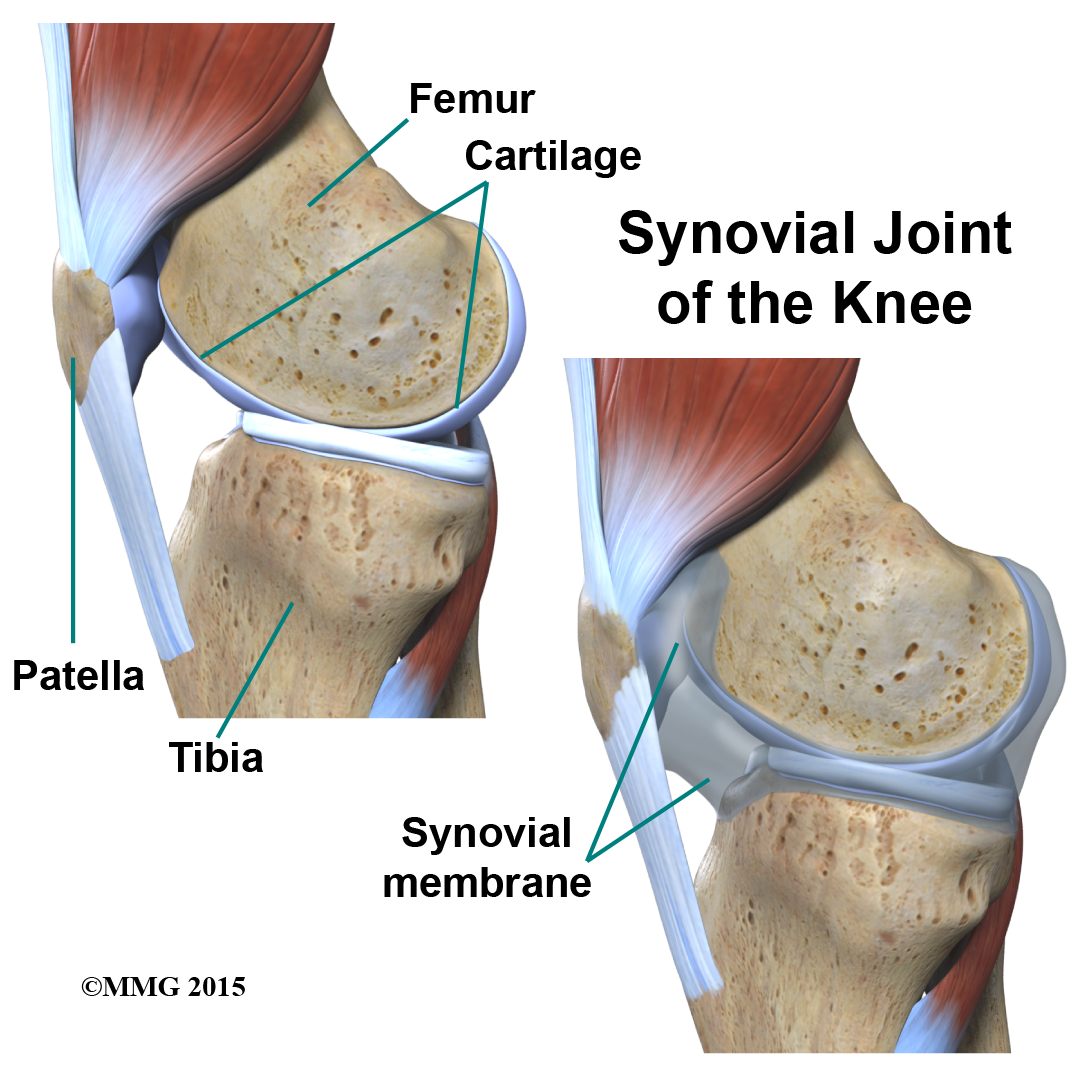
This guide will help you understand what parts make up the knee how the parts of the knee work Important Structures The important parts of the knee include bones and joints ligaments and The knee joint is a synovial joint between the femur, tibia, and patella that allows for flexion, extension, and some rotation, and contains cruciate ligaments, menisci, and surrounding
The anterior cruciate ligament (ACL) is one of a pair of cruciate ligaments (the other being the posterior cruciate ligament) in the human knee. The two ligaments are called „cruciform“ An overview of the anatomy of the knee joint including bony articulations, ligaments, menisci, arterial supply, innervation and relevant
The knee is the biggest joint in your body. It’s also one of the most commonly injured joints. Knees contain bones, cartilage, muscles, ligaments and nerves. This document discusses the anatomy and kinematics of the knee joint. It begins by describing the three bones that make up the knee – the femur, tibia, and patella. It then discusses the
In nearly all circumstances, the knee works in axial compres-sion under the action of gravity. It must therefore reconcile two opposed requirements, namely mobility and stability. This
The static stability of the knee joint complex depends on four major knee ligaments, which provide a primary restraint to abnormal knee motion
- Limpopo — Wikipédia _ Limpopo Südafrika Karte
- Lieutenant-Governor Of British Columbia
- Lilien-Apotheke Gifhorn _ Apotheke Gifhorn Lehmweg
- Liebe American Bully Boxer Welpen Zu Verkaufen Die
- Lily Pad Water Garden Pond A Beginner’S Acrylic Painting Tutorial
- Life Science Vs Medical Science Vs Health Science
- Liebfrauenstraße In Mönchengladbach Neuwerk ⇒ In Das Örtliche
- Life After Death Horror Fest _ Alice Cooper regresa a México; encabezará el Life After Death Horror Fest
- Lilly Hat Lymphdrüsenkrebs _ Puschel-Schilddrüse-Gastritis
- Limerick Junction Station To Mary Immaculate College
- Linde Wiemann Forms Partnership With Indian Jbm Group
- Life On The African Savanna – 21 Interesting Facts About The Savanna