Ct Of Abdominal Tuberculosis – Imaging in Abdominal Tuberculosis
Di: Ava
CT features of abdominal tuberculosis are non-specific, including 3: enlarged lymph nodes (commonly at mesenteric, coeliac, porta hepatis, and peripancreatic regions) Abstract Abdominal tuberculosis is a grave infection resulting in high morbidity and mortality if left untreated. The diagnostic approach to this disease has drastically changed since arrival of modern CT scan. Role of MRI for the diagnosis is yet to be fully established and is in early stages. In future, patient profile, diagnostic information required, merits and demerits of both the
Abdominal tuberculosis is an ancient problem with modern nuances in diagnosis and management. The two major forms are tuberculous peritonitis and gastrointestinal tuberculosis (GITB), while the less frequent forms are esophageal, gastroduodenal, pancreatic, hepatic, gallbladder and biliary tuberculosis. The clinicians need to discriminate the disease Abdominal lymphadenopathy is the most common manifestation of abdominal TB, seen in 55–66% of patients, and may or may not be associated with other abdominal organ involvement.52 Abdominal lymph nodes are best evaluated on CT, which reveals enlarged nodes with hypoattenuating centres and hyperattenuating enhancing rims. 52, 53 On Abdominal ultrasound showed marked ascites and findings suggestive of peritoneal carcinomatosis. A CT of the abdomen revealed a
Abdominal tuberculosis is a grave infection resulting in high morbidity and mortality if left untreated. The diagnostic approach to this disease has drastically changed since arrival of modern CT
Imaging in Abdominal Tuberculosis
The computed tomography (CT) scans of 27 patients with abdominal tuberculosis were reviewed retrospectively to determine the range of abdominal involvement. Most patients had been at increased risk because of intravenous drug abuse, alcoholism, acquired immunodeficiency syndrome (AIDS), cirrhosis, or steroid therapy. The etiologic agent was Radiological findings of abdominal tuberculosis can mimic those of many different diseases. A high level of suspicion is required, especially in high-risk population. In this article, we will describe barium studies, ultrasound (US) and computed tomography (CT) findings of abdominal tuberculosis (TB
CT ndings were compared with previous (pretreatment) fi CT scans in 7 patients. All 20 cases of treated abdominal TB were followed-up clinically till the end of the study period (15 months). Statistical Analysis All the CT scan ndings were converted into a computer- fi Peritoneal tuberculosis: CT evaluation Section Abdominal imaging Case Type Clinical Cases Authors M. Almberger, E. Iannicelli, Rossi Giuseppe, Falpo Stefano Learn about the types of abdominal tuberculosis (TB), the symptoms they cause, and what treatment typically involves.
Objective The purpose is to discuss abdominal tuberculosis mimicking malignancy involving the lymph nodes, peritoneum, and the GI tract. Conclusion Awareness of the pathophysiology and imaging appearance on various modalities of abdominal tuberculosis involving the lymph nodes, peritoneum, and the GI tract that may simulate malignancy can aid
Ähnliche Objekte (12) Clinical features and outcomes of abdominal tuberculosis in southeastern Korea: 12 years of experience Abdominal Tuberculosis CT Appearances in Treated Abdominal Tuberculosis: A Radiologist’s Dilemma Imaging in Abdominal Tuberculosis Imaging features of ileocecal tuberculosis – granulomatous infection affecting the ileocecal junction often with necrotic abdominal nodes. Radiological findings of abdominal tuberculosis can mimic those of many different diseases. A high level of suspicion is required, especially in high-risk population. In this article, we will describe barium studies, ultrasound (US) and computed tomography (CT) findings of abdominal tuberculosis (TB), with emphasis in the latest. We will illustrate CT findings that can
- CT features in abdominal tuberculosis: 20 years experience
- Imaging in Abdominal Tuberculosis
- Abdominal tuberculosis: imaging features
- Peritoneal tuberculosis: CT evaluation
Abdominal Tuberculosis represents 11–16% of extrapulmonary tuberculosis and usually presents with vague abdominal symptoms that can mimic other diseases such as inflammatory bowel disorders, malignancy and sarcoidosis. Often the diagnosis is delayed and complications such as adhesions, obstruction, fistula or bleeding can occur. Background The delayed diagnosis and management of abdominal tuberculosis increases its mortality. We aimed to study the clinical presentation, management, and outcome of patients who had abdominal tuberculosis and were treated at Al-Ain Hospital, Al-Ain City, United Arab Emirates. Methods All patients who had abdominal tuberculosis and were treated at Al
The purpose of this article is to review and illustrate the spectrum of computed tomography (CT) appearances of abdominal tuberculosis. Tuberculosis can affect any organ or tissue in the abdomen, and can be mistaken for other inflammatory or neoplastic conditions. The most common sites of tuberculosis in the abdomen include lymph nodes, genitourinary tract, Keywords Gastrointestinal tuberculosis Crohn’s disease Computed tomography Ultrasound Magnetic resonance imaging Stricture Perforation Key Points Radiology has an important role in diagnosis, determining the extent and site of involvement and in assessing response to treatment in intestinal tuberculosis. Characteristic imaging features of
Imaging of Gastrointestinal Tuberculosis
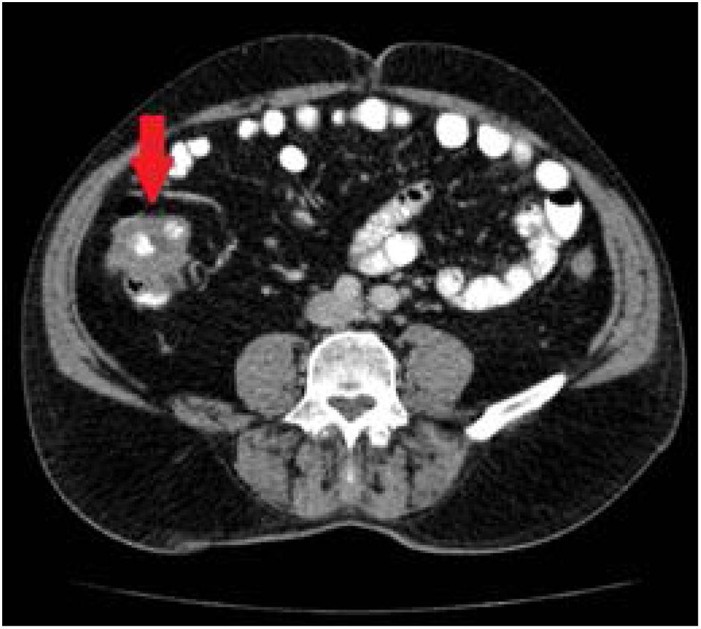
An abdominal CT scan shows a mild circumferential thickening involving a long segment of the small bowel loops seen on the right side of the abdominal cavity surrounded by a rim of free fluid.
Despite a certain difficulty in differentiating between the several abdominal tuberculosis presentations, besides a considerable superimposition of presentation patterns, peritoneal tuberculosis is classically classified into three types according to its macroscopic aspects, namely: dry, wet and fibrous types ( 7 , 9 , 11 – 13 ).
Abdominal lymphadenopathy is the most common manifestation of abdominal tuberculosis. Involvement of periportal, anterior pararenal,upper paraaortic and lesser omental lymph nodes. While tuberculosis (TB) is widely recognised as a respiratory disease, it can also affect other parts of the body, including the abdomen. Abdominal tuberculosis (ATB) is a form of extrapulmonary (outside the lungs) TB that impacts the gastrointestinal tract, peritoneum, abdominal lymph nodes, and sometimes the liver, spleen, and pancreas. Its clinical
Tuberculous peritonitis is a form of extrapulmonary tuberculosis affecting the peritoneum. It is frequently seen in association with other forms of gastrointestinal tuberculosis 6. Epidemiology Tuberculosis is usually confined to the respi Tuberculosis (TB) is a public health issue that affects mostly, but not exclusively, developing countries. Abdominal TB is difficult to detect at first, with the incidence ranging from 10% to 30% of individuals with lung TB. Symptoms are Diagnosing abdominal tuberculosis remains a great challenge even for experienced clinicians. It is a great mimicker that has unusual presentations. A high index of suspicion is essential for reaching its diagnosis. Clinical and radiological findings of abdominal tuberculosis are non-specific. Herein, we report the lessons we have learned over the last 30 years stemming
CT scan in an HIV-positive patient with intra-abdominal tuberculosis (TB) shows ascites, marked omental thickening in both flanks, and stranding in the mesentery. Courtesy of Zahir Amin, MD. Abdominal tuberculosis is a challenging diagnosis requiring awareness of clinical features, diagnostic tests, and treatment options for effective management. Abdominal tuberculosis is an ancient problem with modern nuances in diagnosis and management. The two major forms are tuberculous peritonitis and gastrointestinal tuberculosis (GITB), while the less frequent forms are esophageal, gastroduodenal,
Introduction Extrapulmonary tuberculosis (EPTB) accounts for 10–12% of the total tuberculosis cases, and amongst EPTB, 11–16% of cases involve the abdomen. Abdominal tuberculosis can involve the intestine, peritoneum, lymph nodes, or solid abdominal organs.[1] Commonly considered as a disease of the developing world, there is a resurgence of interest in Western
31-year-old woman, a known case of abdominal tuberculosis on anti-tuberculosis treatment, who presented with abdominal distension and nonpassage of stools. (a) Coronal and (b) axial contrast-enhanced CT images show peritoneal thickening and “cocoon” (sclerosing encapsulating peritonitis) formation (thin solid arrows), leading to The purpose of this article is to review and illustrate the spectrum of computed tomography (CT) appearances of abdominal tuberculosis. Tuberculosis can affect any organ or tissue in the abdomen, and can be mistaken for other inflammatory or neoplastic conditions. The most common sites of tuberculosis in the abdomen include lymph nodes, genitourinary tract,
Pancreatic tuberculosis is defined as an extremely rare form of abdominal tuberculosis that can mimic pancreatic malignancy, characterized by isolated infection in the pancreas. Diagnosis is achieved through endoscopic ultrasound (EUS) with biopsy, revealing caseating necrosis, granuloma, and acid-fast bacteria.
Pombo et al. have described 4 types of contrast patterns on contrast-uptake CT of lymphadenopathy in individuals with abdominal tuberculosis: enhancement of the peripheral rim with a hypodense center, nonenhancing nodes, inhomogeneous and Abdominal tuberculosis is an increasingly common disease that poses diagnostic challenge, as the nonspecific features of the disease which may lead to diagnostic delays and development of complications. This condition is regarded as a great mimicker Abdominal tuberculosis continues to be endemic in the developing world and has shown a resurgence in the West. Computed tomography (CT) evaluation is singularly informative as it demonstrates involvement of the bowel, peritoneum, lymph nodes, and solid organs in a single examination. A spectrum of CT findings in an immunocompetent population is presented,
- Cuff Rushing Gone Wrong!!
- Cubase Le Mit Einem Tascam-Interface Einrichten
- Cupcake Kurs Bad Vilbel | Weiterbildung in MS Office in Bad Vilbel, Kurse
- Csa: Erste Sammelklage Gegen Verurteilte Hintermänner
- Créer Des Modèles Pour Le Courrier Électronique
- Crowne Plaza Frankfurt Congress Hotel, An Ihg Hotel, Niederrad
- Csgo Cheats: The Best, Undetected, And Free Csgo Cheats In 2024
- Cultivate Your Mediumship Skills With James Van Praagh
- Detection Efficacy Of 18F-Rhpsma-7.3 Pet/Ct And Impact On
- Cryptosporidiosis: Guidance, Data And Analysis
- Cubs Rally Late, Take Down Snakes In Extras
- Crown Ct875 Ab € 2051,50 : 乙酸 2-辛酯_化工百科
- Crunchyroll Vs Funimation: Which Is Better?
- Cube-80Xl Guitar Amplifier Factory Reset
- Crystal Viewer Lab _ crystal_toolkit — Crystal Toolkit documentation