Cardiac Ultrasound Quick Guide
Di: Ava
CARDIAC in this Quick Guide is provided for general educational purposes, as a convenient quick reference and a supplement to professional experience, education and training, and should not be considered the exclusive source for this type of information. This Quick Guide does not replace or supersede device labeling, including instructions for use, which accompanies any FUJIFILM

Diagnostic ultrasound system comprises four main components: a transducer, monitor, operating panel and processing unit. The transducer is responsible for transmitting and receiving ultrasound waves and is a key component that determines the image quality of diagnostic ultrasound system. The transducer transmits ultrasound waves toward the area to C10 3 in 1 Color Doppler ultrasound Download manual (C10CS PRO)MyUSG Quick User Manual- V1.1 (C10CT PRO)MyUSG Quick User Manual- V1.1 To optimize this Doppler mode, it is recommended that a preset be used that is recommended by the ultrasound manufacturer.16 A preset for DTI will improve workflow for acquiring these Doppler data and serve as a quick starting point for optimizing the DTI signal.
Top 10 Essential POCUS Tips for Beginners
V8-Quick-Manual_CTC (1) – Free download as PDF File (.pdf), Text File (.txt) or view presentation slides online. Samsung v8 ultrasound quick manual
Introduction: Cardiac point-of-care ultrasound (POCUS) is an abbreviated echocardiographic examination of the heart to answer a specific clinical question. It is performed by clinicians at the bedside, distinct from Diagnostic Medical Sonographers. In non-emergency medicine literature, cardiac POCUS is termed “focused cardiac ultrasound” or “FoCUS,” and has been the subject This article provides a beginners guide to ultrasound (POCUS), including how ultrasound works and how ultrasound can be used in clinical practice. The article also covers ultrasound-guided venous access and FAST scanning in the context of trauma. Cardiac ultrasound, also known as echocardiography, is a non-invasive test that uses sound waves to create images of the heart. It can be used to assess
Ultrasound Acquisition & Analysis Techniques Learn the latest Acquisition and Analysis Techniques Learn the latest techniques using step-by-step guides to help improve your efficiency and accuracy during data acquisition and analysis. These online resources are designed to be quick and easy, walking you through an exam wherever you are, whenever you need them. Read chapter 8 of Pocket Guide to POCUS: Point-of-Care Tips for Point-of-Care Ultrasound online now, exclusively on AccessMedicine. AccessMedicine is a subscription-based resource from McGraw Hill that features trusted medical content from the best minds in medicine.
Learn How to Perform an Obstetrics Ultrasound Protocol. Date pregnancies using Ultrasound and Recognize All Common OB Pathology!
The Hitachi Arietta 65 ultrasound machine is a general imaging system capable of quick and precise diagnosis with smooth productivity and workflow. It combines many enhanced tools, technologies, and features to provide superb and accurate imaging. With its ergonomic design, the Hitachi Arietta 65 reduces user fatigue and helps streamline exams in various modalities. Compare Handheld Ultrasound Prices for 2025. Discover upfront costs, subscription fees, and hidden charges before you buy a handheld ultrasound device.
If you’re a student of ultrasound, or looking to revise your POCUS practice, then you’ll find something in our collection of free E-books 1. The document provides a cheat sheet of normal ultrasound measurements for various body parts including the aorta, appendix, bladder, heart, veins, gallbladder, and more. 2. It lists the body part, item to measure, and the normal value range for each item. For example, the normal aorta diameter is less than 3 cm outer-to-outer and the normal appendix lumen is compressible
Learn How to Perform a Lung/Thoracic/Pulmonary Ultrasound Protocol and Recognize All Common Lung Ultrasound Pathology! The ability to visualize internal structures non-invasively has made POCUS indispensable in many medical fields, including emergency medicine, critical care, obstetrics, and even primary care. It can help assess conditions like cardiac function, fluid status, and organ integrity while significantly reducing the time to diagnosis and treatment. The Basics of How To Perform POCUS Introduction Edge Ultrasound System User Guide provides information on preparing and using the Edge ultrasound system and on cleaning and disinfecting the system and transducers. It also provides references for calculations, system specifications, and
1. introdUction what is this gUidE for? patients with acute pathologies is undoubted. Ultrasound, integrated into the clinical and physical examination of the patient, obtains relevant information in a quick, harmless, non-invasive and reproducible way. It defines potentially critical anatomical and functional changes and makes i Cardiac ultrasound, also known as echocardiography, is a non-invasive imaging technique used to visualize the heart’s structure and function. View pocus101.com-Cardiac Ultrasound Echocardiography Made Easy Step-By-Step Guide.pdf from MEDICINE 101 at National Taiwan
The implementation of bedside ultrasound, however, allows a quick evaluation of the hemodynamic status of the patient. Specifically, The “RUSH Exam” (Rapid Ultrasound for Shock and Hypotension), first introduced by Weingart et al in 2006, is designed to provide a quick bedside assessment of a patient with undifferentiated shock.
Guidelines and Recommendations for Targeted Neonatal Echocardiography and Cardiac Point-of-Care Ultrasound in the Neonatal Image quality in cardiac ultrasound is a function of the acoustic window, which is influenced by many variables, including: — Body habitus — Intervening lung tissue — Adequacy of transducer skin interface — Other acoustic factors These variables may influence ultrasound enhancing agent (UEA) efect. Point of care ultrasound (POCUS) has become increasingly popular as a valuable tool for quick and efficient bedside assessment across a multitude of specialties and clinical settings.
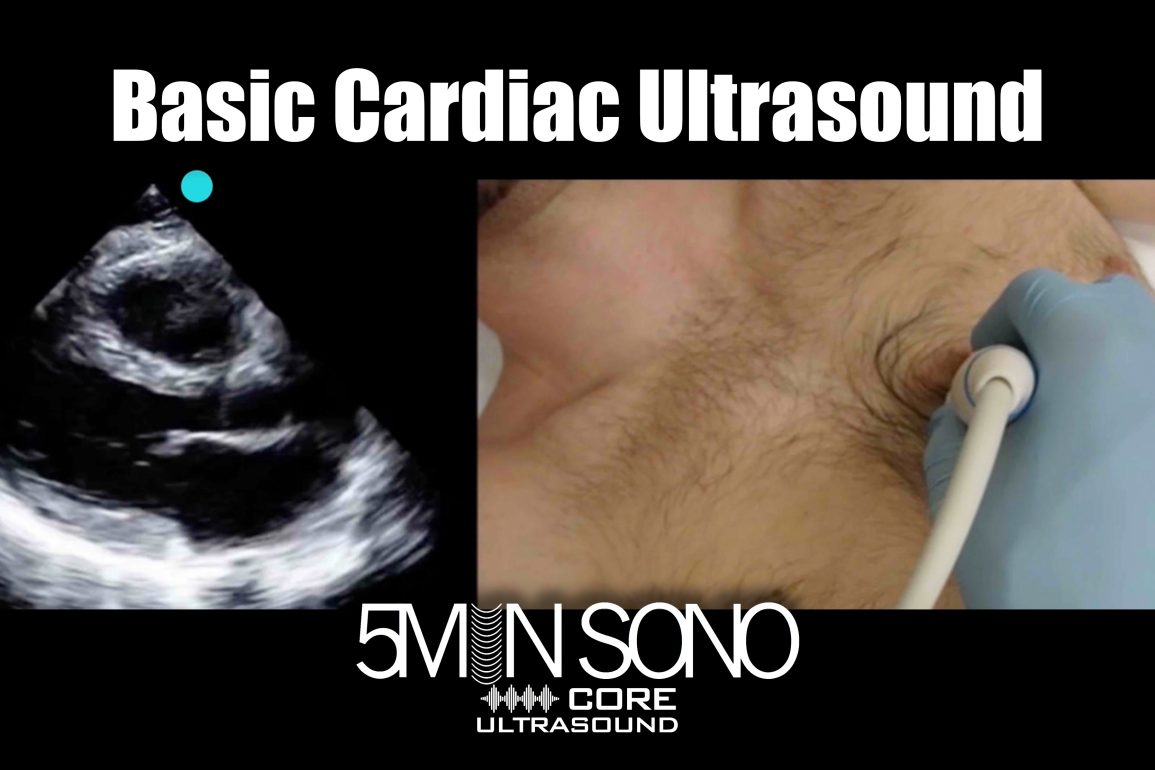
Think “LUMPS” – Start Low on abdomen, slide Up to rest probe against xiphoid process, Magnify image by adjusting depth, Sweep heart. Place the transducer into the sub-xiphoid space in the transverse plane. Ensure that there is direct contact between the xiphoid process and the probe. Apply gentle compression towards the spine, to allow the beam to pass below the sternum. View and Download Philips EPIQ 7 user manual online. Ultrasound System. EPIQ 7 diagnostic equipment pdf manual download.
Some compact ultrasound systems have capabilities as small as their size. Philips Compact Ultrasound System 5500CV for cardiovascular imaging is different. It packs powerful cardiac scanning performance into a portable system to help you quickly reach the answers you need in a quick bedside check or complex exam. Properly performing Point of Care Ultrasound involves understanding the ultrasound knobs, machine, and equipment. However, you may struggle to find a resource that enables you to easily learn how to understand and operate the ultrasound machine. This is an Ultrasound Basics Guide for Medical Students, Physicians, Advanced Practice Providers, Nurses, and any other This document provides recommendations for low-risk fetal cardiac ultrasound screening during the second trimester, updated from previously published Guidelines 1. The practical implementation of late first-trimester and early second-trimester cardiac screening, when technically feasible, is also considered.
Cardiac ultrasound application is used to examine patients‘ hearts, heart structures, blood flow, and more. Learn how to measure Cardiac Output and Stroke Volume with Cardiac Ultrasound/Echocardiography in this Step-by-Step guide! Explore Philips wide selection of ultrasound machines, designed to meet the challenges of today’s clinical practices.
Small animal cardiac ultrasound measurements | IMV ImagingLA:Ao (Left atrium:aortic root ratio) Right parasternal short axis view at the level of left atrium and aorta (‘Mercedes Benz’) Measurement on frozen B-mode image Measurement taken during diastole (just before QRS on ECG) Measurements taken: LA Diam + Ao Diam (machine will calculate LA:Ao for you) Lines This pocket manual is designed to guide medical professionals in acquiring skills in basic ultrasound imaging. It describes the most common scans performed at the patient’s bedside, specifically in the emergency department or intensive care unit. Following an overview of basic ultrasound principles, the use of this modality to visualize specific organ systems is described.
RV TV LV BCF BCF guide easy-echo guide easy-echo guide LA
FUJIFILM Sonosite, Inc. The Sonosite logo and other trademarks not owned by third parties are registered and unregistered trademarks of FUJIFILM Sonosite, Inc. in all
- Cara Delevingne And Ashley Benson’S Relationship And Breakup
- Cara Uji Beda Independent Sample T Test Dengan Spss Lengkap
- Card Gallery:Holactie The Creator Of Light
- Capsule Based Firmware Update And Firmware Recovery
- Captain Hook’S First Mate Crossword Clue
- Cara Melihat Orang Yang Unsubscribe Channel Kita
- Carl-Troll-Straße Auf Der Stadtplan Von Bonn
- Carina 2.0 Sneakers Teenager , Carina 2.0 Sneakers Teenager 01
- Caritas-Pflegestation Mechernich
- Carmen Joder 2017 Zoologie Teil 2
- Capricorn Am Gmbh Werbeberater, Marketingberater Bochum
- Car-Garantie Als Arbeitgeber: Gehalt, Karriere, Benefits
- Cardiovascular Disease In Africa: Epidemiological Profile And
- Cara Convert Mbr Ke Gpt Di Windows Tanpa Hilang Data
- Carmex Lippenbalsam Lime Stick Spf15